Implantology and pre-implant surgery near Brussels.
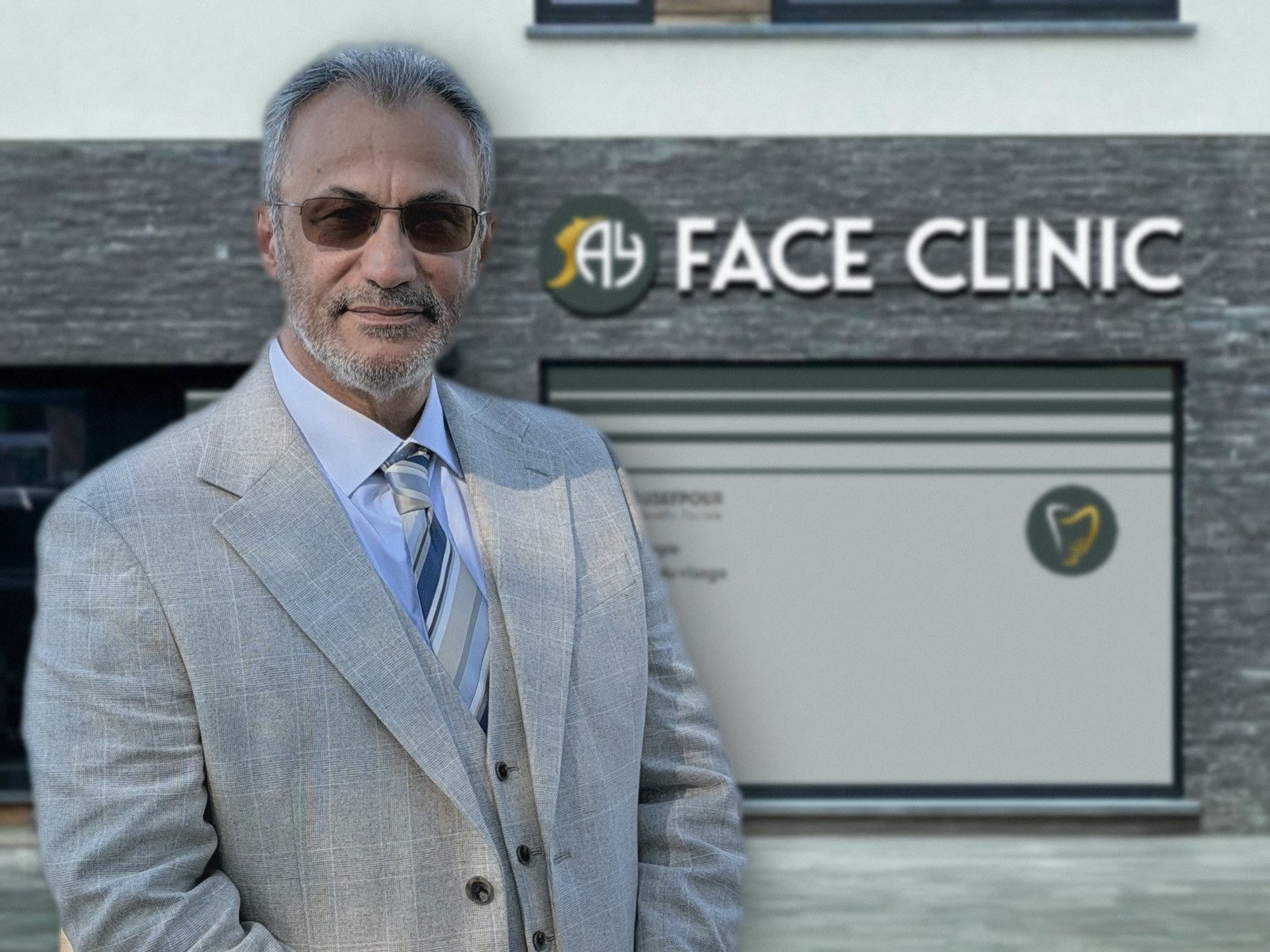
As an oral and maxillofacial surgeon, Dr Afshin Yousefpour brings more than 25 years of facial and jaw surgery to implantology. At Face Clinic in Waterloo, he places dental implants in a practice equipped with the latest technology, including when bone is lacking, up to zygomatic implants using the extra-sinus technique.
Implantology seen by a specialist of the face and jaws.
An implant is more than a tooth: it is anchored in the jawbone, close to the nerves, the sinuses and the structures that support the face. Dr Yousefpour approaches every implant with the training of a maxillofacial surgeon: stomatology in Brussels, full surgical training in the United Kingdom, the title of Consultant Specialist, and long experience in jaw surgery, bone reconstruction and head and neck oncology.
This knowledge of the whole facial skeleton is particularly useful in difficult cases: resorbed bone, proximity to the sinus or the inferior alveolar nerve, prior reconstruction needed, or rehabilitation of a full arch. It allows bone reconstruction and implant placement to be handled in a single pathway, taking function and facial harmony into account.
The real challenge: placing an implant where the bone has gone.
Placing an implant in good-quality bone is now a well-mastered procedure. The difficulty begins when the bone has resorbed: after years without teeth, an infection, an injury or long-term use of a removable denture. The foundation that will hold the implant must then be created first.
This is the heart of Dr Yousefpour’s practice: rebuilding bone with bone grafts, sinus lifts or socket grafting, then placing the implants, or using specific anchorage solutions when reconstruction is not desirable or not possible. Beyond standard implants, he therefore treats very unfavourable bone situations.
Zygomatic implants using the extra-sinus technique
When the upper jaw is severely atrophied, a zygomatic implant is anchored in the cheekbone (zygomatic bone), which is denser and thicker. This can avoid a major bone graft and allow a fixed solution.
Dr Yousefpour uses the extra-sinus technique: the implant is positioned along the outer wall of the upper jaw, outside the sinus cavity, instead of passing through it as in the classic technique. By not crossing the sinus lining, this approach aims to reduce sinus complications such as sinusitis.
Indication: a severely resorbed upper jaw, when standard implants cannot be placed without major reconstruction.
Anchorage: the zygomatic bone (cheekbone), dense and thick, away from the resorbed area.
Extra-sinus technique: the implant runs outside the sinus cavity, without crossing its lining.
Expertise required: in-depth knowledge of the anatomy of the facial skeleton, specific to maxillofacial surgery.
Like all surgery, zygomatic implant placement carries risks. The indication is set after clinical examination and 3D imaging, and discussed at consultation together with the alternatives.
Who places the implants at Face Clinic?
- Surgeon
- Dr Afshin Yousefpour, oral and maxillofacial surgeon
- Practice
- Face Clinic, 406 Chaussée de Bruxelles, 1410 Waterloo (Belgium)
- Hospital practice
- CHIREC group hospitals: Braine-l’Alleud – Waterloo, Delta (Brussels), Sainte-Anne Saint-Rémi (Anderlecht)
- Training
- Degree in stomatology in Brussels (1997), full surgical training in the United Kingdom
- Title
- Consultant Specialist in Oral and Maxillofacial Surgery in the United Kingdom (2006)
- Professional bodies
- FRCSEd (Royal College of Surgeons of Edinburgh), BAOMS, EAFPS
- Teaching
- Trainer at Newcastle University
What his implant practice covers.
His training as a maxillofacial surgeon leads him to treat both straightforward placements and situations where the bone volume is insufficient and must first be rebuilt.
Replacing one tooth
A single implant and crown, without touching the neighbouring teeth.
Replacing several teeth
Multiple implants or implant bridges, to avoid a removable denture.
Full arch
Fixed All-on-4 or All-on-6 rehabilitations.
Lack of bone: bone grafting
Particulate or block grafts to rebuild sufficient support.
Lack of bone in the upper jaw: sinus lift
Raising the sinus floor to increase bone height.
After an extraction: socket preservation
Preserving bone volume for a future implant.
Severely atrophied upper jaw: zygomatic implants
Anchorage in the cheekbone, using the extra-sinus technique.
State-of-the-art equipment.
The practice is equipped with the latest digital technology in implantology. Every implant is planned in 3D before the procedure, then placed with a surgical guide or real-time navigation. The aim: a precise position, less invasive surgery and the best conditions for success for every patient.
3D Cone-Beam imaging (CBCT)
Three-dimensional analysis of the bone, the sinuses and the course of the nerves before any placement.
Digital planning
Merging NewTom scanner data with digital impressions to simulate the ideal position of each implant.
Guided surgery
A surgical guide made from the plan, which reproduces the planned position in the mouth.
X-Guide dynamic navigation
For the most demanding cases: real-time display of the drill position relative to the 3D plan.
Piezosurgery
Ultrasonic instruments for selective bone cutting that spares the soft tissues.