
Bone grafting
Expert bone reconstruction before implant placement.
Duration
45 minutes to 2 hours
Anaesthesia
Local, with advanced comfort protocols
Hospital stay
Day case
Recovery
Swelling peaks at 48 to 72 hours, settling within about ten days
Dr Yousefpour, oral and maxillofacial surgeon in Waterloo, offers expert management of bone atrophy. Combining scientific rigour, digital planning and advanced surgical techniques, our mission is to restore lost anatomy lastingly to ensure the success of your implant treatment.
“My role as a surgeon is to turn a compromised site into fertile ground. Thanks to guided surgery and digital technology, every reconstruction is designed to give lasting implant stability.” — Dr Yousefpour
The protocol, step by step.
- Step 1
Diagnosis and 3D analysis
Detailed study of bone density and volume on the CBCT.
- Step 2
Digital simulation
Creation of your personalised surgical guides for optimal precision.
- Step 3
Expert surgery
Procedure performed under advanced comfort protocols.
- Step 4
Post-operative follow-up
Careful support for optimal healing.
Socket preservation: the foundation of a lasting result
Preserving bone after extraction. When a tooth has to be removed, the bone naturally starts to resorb. To prevent this loss of volume, we routinely perform socket preservation.
High-quality bone substitutes: we use purified bone matrices of bovine or porcine origin, recognised for their biocompatibility and their ability to act as a scaffold for your own bone regeneration.
Protective barrier membrane: membranes are essential. Whether resorbable (collagen) or, in more complex cases, non-resorbable, they act as a shield. By preventing soft-tissue invasion, they allow bone regeneration to take place in a protected, stable environment.
Advanced surgery: autologous block grafts and GBR
Three-dimensional reconstruction for major bone loss. When the remaining volume is insufficient, we offer complex reconstruction protocols, favouring the “gold standard”: the autograft.
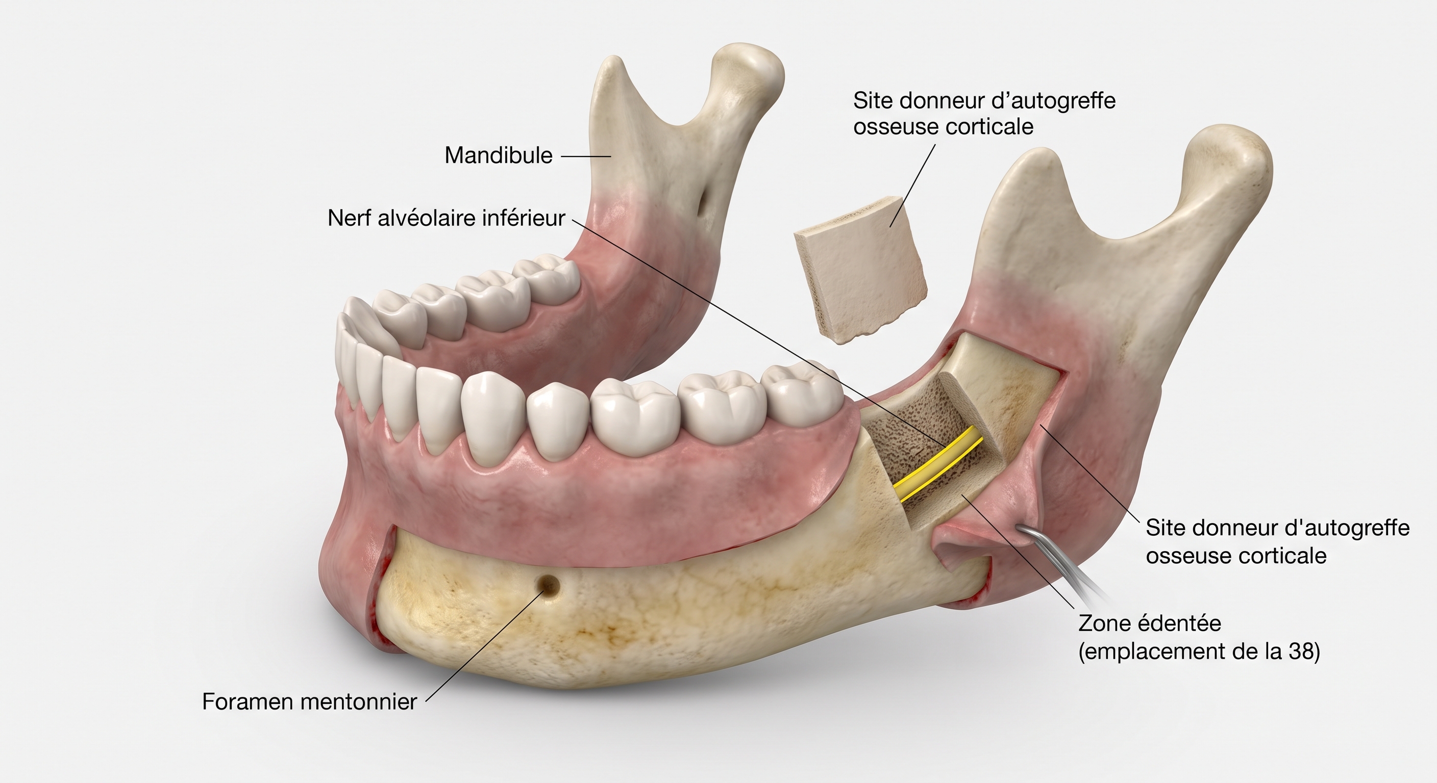
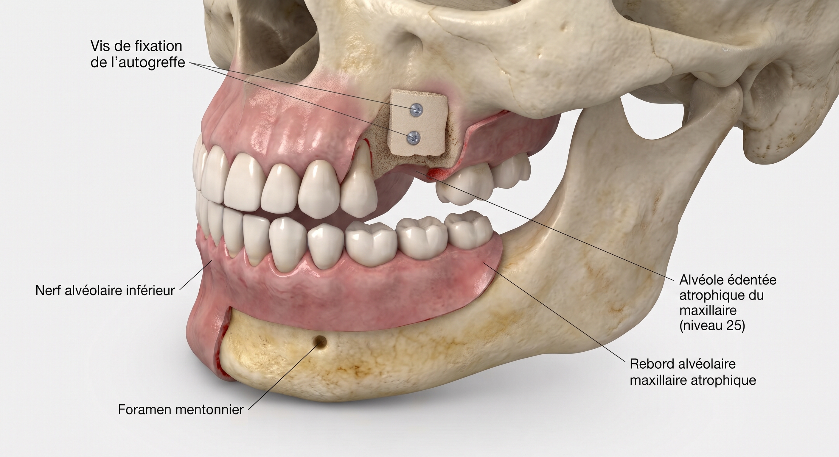
Autologous block grafts: using your own bone tissue is the most predictable technique. These grafts, taken from donor sites inside the mouth, integrate optimally and can rebuild severe horizontal and vertical defects.
3D guided bone regeneration (GBR): for complex cases, we combine this expertise with GBR techniques using titanium meshes or custom reinforced membranes. These devices maintain a dedicated space for reconstruction, ensuring an ideal bone architecture for your future implants.
Precision through guided surgery (mandibular angle)
The major innovation in our practice lies in optimising bone harvesting at the angle of the lower jaw.
3D planning: from your CBCT imaging, we analyse the anatomy of the lower jaw in detail, precisely locating the inferior alveolar nerve and other key structures.
Custom harvesting guides: we design and print specific surgical guides that make the procedure safer. This technology allows millimetre-precise bone cuts, considerably increasing the safety of the harvest, reducing operating time and optimising the quality of the bone block obtained.
Why guided surgery? It turns a complex operation into a standardised, highly safe and predictable procedure.
Post-operative protocol: general recommendations and comfort
Pain management: a suitable prescription (painkillers and, if needed, anti-inflammatories or antibiotics) is given at the end of the procedure. Follow the dosages carefully.
Swelling: swelling is a normal reaction after bone surgery. It usually peaks between the 48th and 72nd hour.
Cold: apply ice packs (wrapped in a cloth, never directly on the skin) to the operated area intermittently (15 minutes on, 15 minutes off) for the first 48 hours.
Bleeding: slight pinkish oozing is common in the first 24 hours. If bleeding is heavier, bite gently but firmly on a sterile gauze for 30 minutes.
Oral hygiene
Brushing: brush your teeth regularly but strictly avoid the operated area for the first few days, so as not to disturb healing or strain the stitches.
Mouthwash: use no mouthwash on the day of surgery. From the next day, use the prescribed antiseptic solution gently, without vigorous mouth movements (preferably “passive” rinsing).
Strictly avoid: spitting, sucking on the operated area and touching the stitches with your tongue or fingers.
Diet and lifestyle
Diet: eat soft, cold (or lukewarm) food for the first 4 to 5 days. Avoid hot, spicy or acidic food and food with small seeds (strawberries, sesame, etc.) that could lodge in the wound.
Smoking: tobacco is a major enemy of bone healing and significantly increases the risk of graft failure. Stopping completely or cutting down drastically is essential throughout the consolidation phase.
Physical activity: avoid strenuous effort, heavy lifting and sport for at least one to two weeks.
Information and consent: nature of the procedure and options
You have insufficient bone volume in your jaws, which as it stands does not allow safe and lasting placement of dental implants. To address this, a (simple or complex) bone reconstruction or augmentation may be proposed. Depending on your clinical situation, the procedure may involve:
Socket preservation or a simple graft: filling extraction sockets with purified bone substitutes (of bovine or porcine origin) combined with a protective membrane (resorbable or not).
Complex surgery with an autologous block graft: harvesting your own bone (notably at the angle of the lower jaw, optimised by digital planning and custom guided surgery) to correct major three-dimensional defects.
3D guided bone regeneration (GBR): using meshes or reinforced membranes to maintain the reconstruction space.
Alternatives: no treatment (not replacing the missing teeth), a dental bridge (which involves drilling down healthy teeth) or a removable denture (less comfortable).
Usual recovery
Swelling: common in the cheek, peaking between 48 and 72 hours and settling within about ten days. Cold and following the prescriptions help limit it.
Bruising: bruises may appear on the skin or gums.
Pain: usually moderate and well controlled with the prescribed painkillers.
Temporary limitations: temporary difficulty opening the mouth or slight bleeding in the first 24 hours are possible.
Potential risks and complications
Despite all the care and expertise applied, every operation carries risks. The following complications, some of them rare, may occur:
Wound infection or graft rejection: may require additional antibiotics, or even partial or complete removal of the grafted material.
Early exposure of the membrane or graft: requires close monitoring and sometimes a change in healing strategy.
Altered sensation (paraesthesia): because of the proximity of the inferior alveolar or mental nerve, numbness, tingling or temporary (more rarely permanent) loss of sensation of the lip, chin or teeth may occur.
Sinusitis or oro-antral communication: for procedures involving the upper jaw (sinus lift).
Partial or complete graft failure: could compromise future implant placement and require further reconstruction.
Aggravating factors and patient commitments
Smoking: tobacco severely impairs microcirculation and greatly increases the risk of graft failure and implant loss. Stopping or cutting down drastically is strictly required.
Hygiene and compliance: strict adherence to the post-operative instructions (suitable diet, careful hygiene, not touching the area) and attendance at all follow-up appointments.
General health: accurate and complete information for Dr Yousefpour on your medical history, allergies and current treatments (particularly anticoagulants or bisphosphonates).
Informed consent
Before the procedure, you receive an information and informed consent form at Face Clinic in Waterloo. By signing it, you confirm that you have received all the necessary explanations about the nature of the bone graft, its advantages, alternatives and risks, that you have been able to ask all your questions, that you understand no absolute guarantee of success can be given in surgery, and you authorise the surgeon to adapt the surgical strategy during the operation if medical reasons require it.
Frequently asked questions
Why a bone graft before an implant?
An implant needs enough bone volume to be anchored securely and lastingly. After a tooth is lost, the bone gradually resorbs. The graft rebuilds this lost volume to give the implant a solid foundation.
Where does the grafted bone come from?
It can be your own bone (autograft), for example taken from the ramus of the lower jaw, as particles or blocks, or a bone substitute. The choice depends on the volume to rebuild and your situation.
Is bone grafting painful?
The procedure is performed under local anaesthesia with advanced comfort protocols, as a day case. Swelling peaks between 48 and 72 hours, then settles within about ten days.
How long before the implant can be placed?
A period of bone maturation is needed, usually 3 to 6 months depending on the case, so that the implant is placed in regenerated, solid and stable bone.
Why entrust bone reconstruction to a maxillofacial surgeon?
Rebuilding jaw bone requires knowledge of the whole anatomy of the site: the course of the inferior alveolar nerve, the mental foramen and the maxillary sinus. Dr Yousefpour, oral and maxillofacial surgeon, treats bone atrophy including severe cases, with 3D planning and custom surgical guides.